

3D composition imaging by extreme ultraviolet, laser ablation, mass spectrometry
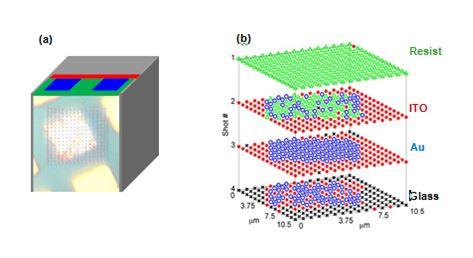
3D composition imaging by extreme ultraviolet, laser ablation, mass spectrometry. Figure (a) sample consisted of 8 mm wide Au pillars 30 nm thick fabricated onto an indium-tin-oxide layer (ITO) 20 nm thick deposited onto a glass substrate. A polymer resist layer 50 nm thick planarized the sample. Figure (b) depicts composition map obtained from the analysis of mass spectra from four consecutive single-shot laser ablation events at each 16x16 grid of probed sites. The ion image plots the distribution resist, ITO, Au, and glass versus laser shot number.
Related center: