

CELL-MET5-I-fig2
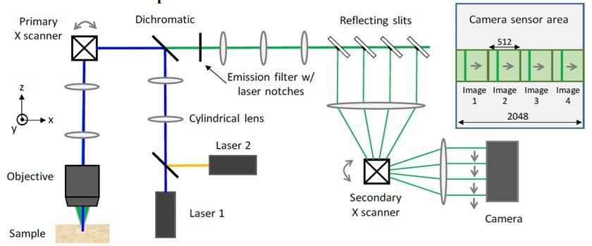
Figure 2. Line-scan Multi-Z confocal microscope. Lasers are formed into a line with a cylindrical lens. Line is scanned along X with primary scanner, Resultant fluorescence is de-scanned and then re-scanned with secondary scanner after being filtered with reflecting slits. Multi-Z images are then projected onto a single camera. Four sections shown in schematic and inset of camera sensor. Allowances can be made for up to 8 sections per camera frame.
Credit:
CELL-MET
Related content:
Related center: